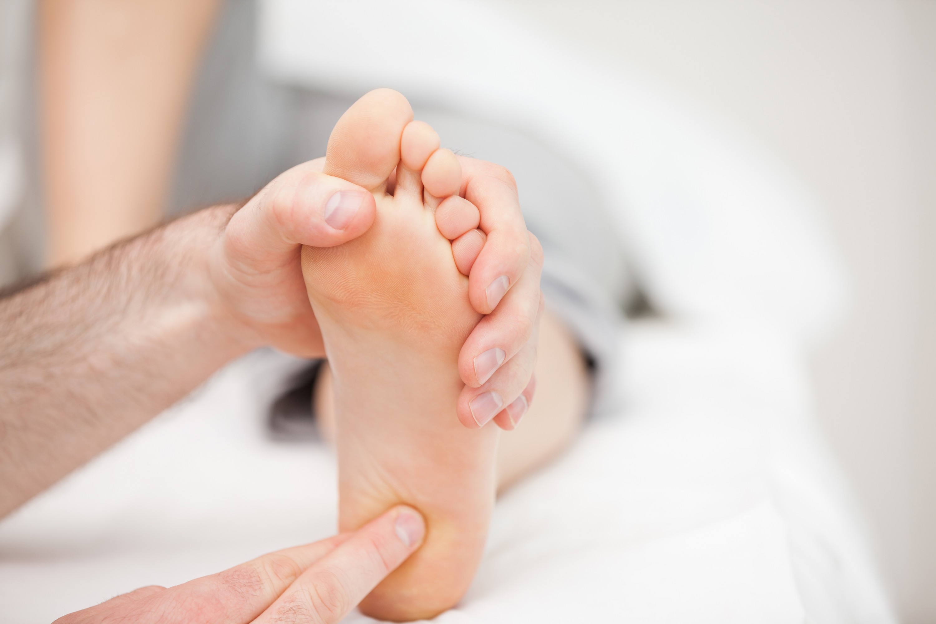
Plantar Fasciitis
Key Points
- Estimated 10% of the population will experience plantar fasciitis over the course of their lifetime;
- Pes cavus and pes planus foot types are at high risk;
- < 10 degrees of ankle dorsiflexion a contributing factor;
- Leg length discrepancy (LLD) also a contributing risk factor;
- Accommodate for heel spurs, if present, in CFO design and footwear modifications.
Definition
- A painful localized syndrome affecting the plantar fascia or plantar aponeurosis, a thick fibrous band of connective tissue that originates at the medial tubercle of the plantar calcaneus and inserts into the plantar plates of the metatarsophalangeal joints, spanning the medial longitudinal arch;
- A degenerative syndrome caused by repeated microtrauma to the plantar fascia.
Pathology
Etiology not clearly understood but mechanical stress and microtrauma associated with a variety of factors;
- Weight-gain;
- Work related acitivity (prolonged weight-bearing);
- Anatomical factors, pes planus or pes cavus;
- Reduced ankle dorsiflexion;
- Leg length discrepancy;
- Excessive lateral tibial torsion;
- Excessive femoral anteversion;
- Overtraining in athletic individuals;
- Reduced muscle strength leading to adult acquired pes planus in the elderly.
Patient Perspective
- localized plantar calcaneal pain upon first rising in the morning or after a period of non-weight-bearing
- symptoms can be bilateral but are frequently unilateral
- pain frequently diminishes after initial rising but returns after prolonged weight-bearing activity
- tenderness on the antero-medial aspect of the inferior calcaneus but may migrate to medial, central and lateral bands of the plantar fascia
- 80% of population experience reduced ankle dorsiflextion
- patient may present with antalgic or altered gait pattern in an attempt to avoid the pain
- bone spurs may be identified by radiographic or ultrasonographic imaging
Differential Diagnosis
If patient presents with symptoms of ???, also consider the following conditions:
- Calcaneal stress fracture;
- Peripheral nerve entrapment;
- Nocturnal pain;
- Tarsal tunnel syndrome;
- Sever’s disease;
- Reiter’s syndrome;
- Ankylosing spondylitis;
- Rheumatoid arthritis;
- Psoriatic arthritis;
- Juvenile idiopathic arthritis (JIA);
- Tumors or fibromas;
- Infection.
Common Testing
- Patients main complaint is pain in the antero-medial subcalcaneal
- A detailed history is required to rule out misdiagnosis
- If bone spurs are present, confirmed by radiographic imaging or ultrasound, take these into account.
Biomechanical tests recommended:
- NWB –> palapte medial tubercle of calcaneus –> note pain;
- NWB –> gently dorsiflex toes to elongate plantar fascia and palpate distally medial, central and lateral bands –> note nodules and pain;
- Evaluate open chain passive ankle dorsiflexion in subtalar neutral –> less than 10 degrees significant –> palpate gastrocnemius and soleus to confirm/rule out isolated contractures;
- Evaluate closed chain biomechanics and gait for excessive pronation/supination. Deal with these in the CFO design and footwear modifications to reduce strain on the plantar fascia and antero-medial calcaneus;
- Check for asymetrical gait pattern –> is LLD present;
- If LLD, assess further.
Contraindications
- Minimize prolonged weight-bearing activity;
- Eliminate barefoot walking on hard surfaces until symptoms subside;
- Minimize activity level, especially intense high-impact activities, and especially for athletic individuals;
- Avoid heel spur and heel pads where loss of sensation is present such as in the diabetic population.
Common Treatment
- Custom foot orthoses to address STJ pronation or supination;
- Heel plugs, holes, horseshoe/spur accommodations for cases where heel spurs are present (consider these carefully in the case of diabetics where neuropathy is present);
- Dorsiflexion night splint or Strassburg sock to keep plantar fascia, gastrocnemius, and soleus in elongated position to improve passive ankle dorsiflextion;
- Lifts, if LLD;
- Appropriate footwear;
- Indoor footwear (especially to avoid barefoot walking);
- Plantar fascia, gastrocnemius, soleus, and foot specific strengthening exercises.
Outside Treatments (outside pedorthis scope of practice):
- Icing;
- Rest/reduction of weight bearing activity;
- Taping;
- Anti-inflammatory medication;
- Physiotherapy;
- Registered Massage Therapy;
- Chiropractic;
- Cortisone or steroid injections;
- Extracorporeal Shockwave Therapy;
- Ultrasound guided dextrose injections (prolotherapy);
- Surgical intervention: fascial release, fasciotomy.
Source: 2012 Clinical Practice Guidelines, Pedorthic Association of Canada


Post This!
Share this post with your friends!